Dr. Shuvo Dutta

Senior Consultant Cardiologist

MD(Cal), MRCP(UK), FRCP(London), FACC(USA)

Director – Interventional Cardiology,
Head Quality Assurance – Cathlab
B. M. Birla Heart Research Centre, Kolkata

Angiography

Coronary Angiography

Coronary angiography is a test that uses dye and special x-rays to show the insides of your coronary arteries – the arteries which supply oxygen-rich blood to your heart.
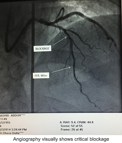
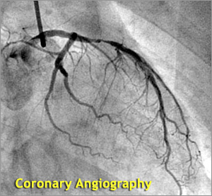
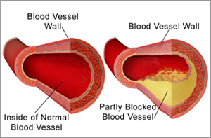
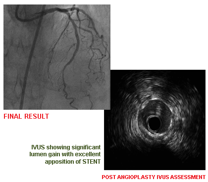
In Coronary angiography, a contrast material (dye) that can be seen using x-ray equipment is injected into the blood vessels of the heart to identify blockages and till now remains the Gold Standard Test. This allows your health care provider to view the flow of blood through your heart.

How is the Test Performed?

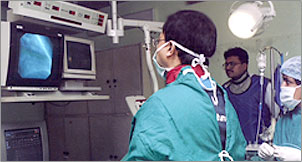
After administering a mild sedative, an area of your body (the arm or groin) is cleaned and numbed with a local anesthetic. The cardiologist passes a thin hollow tube, called a catheter, through an artery and carefully moves it up into the heart. X-ray images help the doctor position the catheter.
Once the catheter is in place, dye (contrast material) is injected into the catheter. X-ray images are taken to see how the dye moves through the artery. The dye helps highlight any blockages in blood flow.
The procedure may last 5 to 10 minutes. You should not eat or drink anything for 8 hours before the test starts. Tell your doctor if you are allergic to seafood, if you have had a bad reaction to contrast material in the past, if you are taking Viagra, or if you might be pregnant.

Peripheral Angiography

A peripheral angiogram is a diagnostic study that uses X-rays and dye to help your doctor detect blockages in arteries other than the heart, viz. those arteries that supply blood to your legs. The test is also called a Peripheral Arteriogram.

IVUS (Intravascular Ultrasound)

IVUS (OVERVIEW)

Intravascular Ultrasound (IVUS) is a medical imaging technology that allows cardiologists to obtain images of diseased blood vessels from inside the artery, using sound waves

Why IVUS?

Evolving changes for better long-term results of Coronary Angioplasty has evolved by help of newer devices which help access vessels to be stented.
A specially designed thin tube with a miniaturized ultrasound detector which sends and receives waves from the vessel walls and translates it into images. This helps to identify the nature of blockage material and helps cardiologists decide strategy for Angioplasty.
After complex Angioplasty, satisfactory stent placement can also be verified using this technique.

FFR

Fractional Flow Reserve

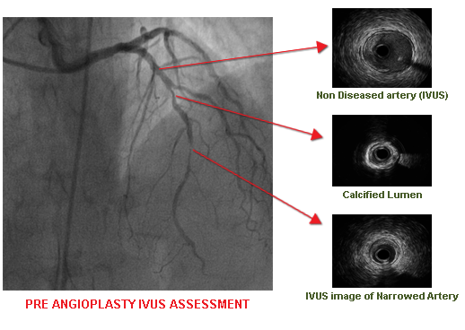
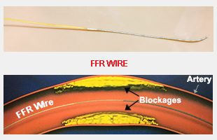
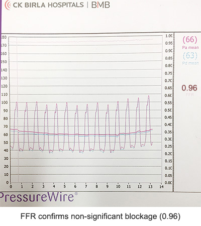
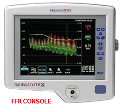
FFR (Fractional Flow Reserve) is a fine wire which measures pressure within the blocked artery before and after the blockage and hence calculates automatically the significance of the blockage.

a) < 0.8 – Significant blockage, needs treatment

b) > 0.8 – Non-significant

Get in Touch

Contact Info

+91 98305 06845

Mobile (Secretary)

shuvodutta@yahoo.com

Doctor's Email

Inquiry Form